|
Xuzhou Ruihua Electronic Science&Technology co.,ltd
|

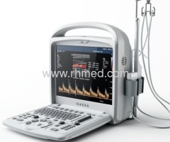
4D Color Ultrasound Scanner
| Payment Terms: | T/T, |
| Place of Origin: | Jiangsu, China (Mainland) |
|
|
|
| Add to My Favorites | |
| HiSupplier Escrow |
Product Detail
4D(Real-time 3D)
4D, or the Real-time 3D technology is another major breakthrough Ruihua has achieved.
A powerful Color Doppler System
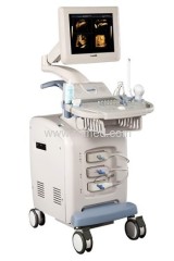
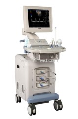
EXRH-800 DC is the 1st general purpose full-digital color Doppler ultrasound diagnostic system. Ruihua Electronic Science & Technology CO.,LTD launched to global market. Carrying 100% China self-owned intellenctual property, the development of EXRH-800 DC made a key project in China's 11th's Five-year Technology Development Plan and made headline of the most prestigious CCTV(China Central Television Station) News in March, 2007. Based on Ruihua's leading full-digital ultrasound imaging technology and image processing technology, ECRH-800 DC features superb image ergonomic design advanced hardware platform, powerful software function and professional clinical application. In particular, EXRH-800 DC is capable of 4D imaging.
4D, or the Real-time 3D technology is another major breakthrough Ruihua has achieved. Through the 4D module that is embedded in the ultrasound system, EXRH-800 DC could easily realize real-time 3D imaging functionalities such as driving of 3D volumetric probe, 3D data acquisition, 3D data reconstruction and post-processing. Apart from being a useful aid to diagnosing neonatal defects, the 4D package enables live display of fetus inside the mother's body, which remains a forever pleasure to the mother and the family.
Complete Clinical Applications:
CFM(Color Flow Mapping)
PW(Pulse Wave Doppler)
CDE(Color Doppler Energy)
Dlr. CDE(Directional CDE)
HPRE(High Pulse Repetitive Frequency)
Triplex(B+CFM+PW)
3D/4D
Extended Field of View Imaging
THI(Two groups of harmonic wave on each probe)
SRI(Speckle Reduction Imaging)
Intraoperative Ultrasound
Ultrasound Guided Biopsy
Superb phased-array cardiac imaging and professional cardiac measurement software package
Unique Clinical Functionalities
RTNT(Real-time network transfer of bother images and videos)
SWR(Video recording up to 1hr).
Direct Printing(Freeze-Print, bypassing workstation or DICOM. Save time and cost of thermal paper)
Support all printers(End-users could install drive for any video, inkjet and laser printers themselves)
One click restoration of O/S
Internally embedded workstation(Support multiple templates and expert annotation)
Automatic identification of probes.
Flexible OB formula (Obstetric formula could be defined and revised by end-users)
Integrated super volume HDD, DVD-RW and dual USB storage.
Continuously upgradeable
Voice command of operation
High-precision Continuous Digital Beam-former
Fine ultrasound beam effectively abates sidelobe noises, significantly improves space resolution and contrast resolution and clearly shows scanned structures in the whole field.
Dynamic Frequency Integration Imaging Technology
Auto-adaptively controls the transmitting and receiving frequency from the near field to the far field and leads to perfect integration of powerful penetration and fine resolution
High-precision Dynamic Receiving Focus
Hi-precision dynamic receiving focus on the whole field gives genuine and fine tissue information
Super Wide-band Imaging Technology
Optimal central frequency selectable based on people characteristics
Self-adaptive Image Optimization Processing Technology
Automatically optimize digital parameters based on current tissue information to display better images
Multi-beam Imaging Technology
Transmit and receive multiple beam at one time to bring clear and authentic images
Self-adaptive Vascular Imaging Technology
Automatically adopt appropriate filtering plan to abate noises of the received Doppler echoes to reach perfect combination of resolution and sensitivity and brings better blood images
Self-adaptive Doppler Imaging Technology
Magnify weak Doppler signals and strengthen pulse wave signals through complicated digital processing to improve both Doppler sensitivity and display
THI (Tissue Harmonic Imaging) Technology
Transmit ultrasound signals at low frequency but form images with high-frequency harmonic signals in echoes to guarantee deep penetration, enhance resolution and eliminate sidelobes
Precise Wall Filer
Wall filter divided into more and finer steps to enhance image quality
Anatomical M Mode Imaging
Sampling line 360°rotatable around any point of the line
Automatic Identification of Probes
Probes are automatically identified, with all parameters preset to the optimal for the selected clinical application.
Related Search
Ultrasound Scanner
Diagnostic Ultrasound Scanner
Veterinary Ultrasound Scanner
Digital Ultrasound Scanner
Ultrasound Scanner Portable
Convex Ultrasound Scanner
More>>
Find more related products in following catalogs on Hisupplier.com
Company Info
Xuzhou Ruihua Electronic Science&Technology co.,ltd [China (Mainland)]
Business Type:Manufacturer, Trading Company
City: Xuzhou
Province/State: Jiangsu
Country/Region: China (Mainland)
|
Jason:
|
|
Jason:
|